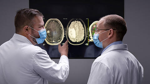
People who develop symptoms often go to their primary care physician or go to an emergency department or primary care doctor. For these people, imaging is the next step with a CT scan if the head or MRI of the brain. The MRI provides more detail that gives physicians a good estimate of the type of brain tumor they are seeing and if it is benign or malignant. You will need a biopsy (obtaining tissue through surgery) for a definitive diagnosis by a neuropathologist.
Treating Primary Gliomas and Meningiomas
Our team approach starts from the moment you arrive at one of our Cancer Network locations. In our tumor board — a weekly gathering of neurosurgeons, neuro-oncologists, radiation oncologists, neuroradiologists, neuropathologists, neuropsychologists, researchers — all of our specialists weigh in on a recommended treatment plan based on your individual situation. By the time we meet, we have heard your story and have seen imaging from your initial evaluation. We feel like we know you already.
When our team treats your glioma or meningioma, you will find a highly personalized approach, a must for brain tumors. We adjust treatment as needed based on your unique response as treatment proceeds. During treatment and follow-up, any team member is available to answer questions and address your concerns.
Your team will often start you on steroids right away. You are often started on anti-seizure medication — even if you don’t have seizures — as some people experience seizures in the period between diagnosis and surgery. You may be able to discontinue steroids and/or antiseizure medications in the future, if you no longer need them.
Some small, slow-growing meningioma that aren’t causing symptoms may not need immediate treatment, but can be carefully monitored with periodic imaging. If the tumor remains stable, you may never need treatment.
Surgery to Remove the Tumor and Preserve Brain Function
For gliomas and meningiomas, your team will talk with you about surgery to alleviate symptoms, reduce pressure on the brain and remove as much of the tumor as is safely possible. They may also discuss collecting tissue that will reveal your tumor type and its grade.
We may also perform surgery to alleviate symptoms like headache, nausea or blurred vision, or to reduce the tumor’s size before radiation therapy or chemotherapy if those therapies are part of your treatment plan. Some tumors cannot be treated with surgery because they have spread too widely into critical brain areas, including the brain stem. The brain stem is a small but critically important part of your central nervous system. Nerve connections from the brain’s cortex move through the brain stem to communicate with your peripheral nervous system, regulating heart and breathing function, consciousness and sleep.
Good functional outcomes are always our priority. Our surgeons specialize in state-of-the-art techniques to keep you safe while removing as much tumor as possible. This has been shown to give patients the best possible outcome. However, if the surgery can’t be done without causing significant deficits in your ability to think, speak, understand and move, we will consider other treatments. Current studies show that survival is best when we remove as much tumor as possible while preserving a patient’s faculties – the qualities that let people be themselves.
Before Your Surgery
Surgically removing a brain tumor often requires cutting the blood vessels that supply the tumor. This can lead to significant bleeding. To help reduce bleeding during surgery, interventional neurologists often perform pre-operative tumor embolization, a procedure that cuts off the tumor blood supply prior to tumor removal.
Tumor embolization can be done the day before surgery or the morning of the operation. We start by identifying the blood vessels that supply the tumor using a catheter device and imaging guidance. Once identified, the interventional neurologist injects micro-particles into the key tumor blood vessels. The particles clump together, forming a dam that cuts off the blood supply to the tumor. During a typical procedure, the interventionalist will embolize from two to six arteries. The goal is to de-vascularize as much of the tumor as possible.
Cutting off a tumor’s blood supply results in a safer tumor removal surgery with less blood loss and a shorter operation time. Embolization often enables surgeons to remove more tumor tissue with less risk.
Brain Tumor Surgery Techniques
In some cases, we are one of only a few programs in the Midwest offering these cutting-edge techniques. You have options, and your care team will work with you to determine the best course of action for your particular brain tumor and symptoms.
Our fellowship-trained experts specialize in brain tumor surgery at the only academic medical center in southeastern Wisconsin. Some of our neurosurgeons treat brain tumors and nothing else. We are constantly looking for ways to improve brain tumor survival rates through research and clinical trials.
-
Microsurgery
Our surgeons are trained in the most advanced microsurgical techniques to remove as much tumor as possible while keeping normal brain structures intact. When a persons condition requires it, surgery is done under an operative microscope to improve the surgeon’s ability to see and protect microanatomy.
-
Laser Interstitial Thermal Therapy (LITT)
LITT is a relatively new minimally invasive option for some brain tumor patients, particularly those with smaller primary brain tumors and metastases. The tumor needs to be less than 3 cm, and the patient cannot have an excess of fluid buildup in the brain (mass effect or edema).
LITT is less invasive than open craniotomies where a large part of the skull is removed. It involves “ablating” rather than resecting tumor tissue. Using this technique, the surgeon makes a tiny incision (less than .5 cm) through which a laser probe is passed into the tumor. The patient is then brought to the MRI scanner with the laser probe in place, and MR-thermography (real-time heat mapping) is used to monitor the laser’s delivery of heat to the tumor. Delivered over a short time, the heat kills tumor tissue.
This procedure has been shown to decrease wound-healing complications and length of stay in the hospital when compared to a traditional craniotomy procedure. Most patients do not need to stay in the ICU after surgery. Depending on the type and location of tumor treated, patients can often go home one or two days after the procedure. Most patients do not need other cancer treatment therapies after having LITT.
In very rare cases (less than 1%), there is a risk of bleeding during the biopsy or laser placement. We have procedures in place to reduce this risk.
-
Fluorescent Brain Tumor Surgery, 5-ALA

Max Krucoff, MD, neurosurgeon, uses fluorescence guidance to get a clear picture of the brain tumor he is removing. We always want to preserve as much healthy brain as possible. Fluorescent brain surgery, or fluorescence-guided surgery (FGS), highlights the tumor cells — giving the surgeon a clear view of what needs to be removed. If the surgeon is able to remove all of the enhanced tumor without injuring too much normal brain, this has been shown to increase life expectancy and quality of life.
Patients undergoing an open brain tumor surgery (craniotomy) for a high-grade glioma are candidates for FGS.
On the day before or the morning of your surgery, you will drink the "Pink Drink" — 5-ALA (5-aminolevulinic acid). It is white powder dissolved in water. We call it the "Pink Drink" because it goes through your system, crossing the blood brain barrier and infiltrating malignant brain cells — making them glow pink or red under a special blue light. 5-ALA can be used in any tumor that "enhances" on an MRI scan, such as glioblastomas and other malignant brain tumors.
Other than the drink and the blue light, the surgery progresses like other open brain tumor surgeries (craniotomies). Side effects specific to the 5-ALA compound include a sensitivity to bright light. Your care team will make sure you are protected from sunlight or bright indoor lighting before and after surgery, and you should avoid strong sunlight for two weeks.
You may experience nausea, anemia (low iron), low platelets, elevated white blood cell counts and temporary liver problems from the combined effects of the surgery, the 5-ALA and the anesthetic. All surgeries carry the risk of blood clots, and brain surgery has a risk of neurological disorders. Talk to your care team about what to watch for and when to alert them regarding any issues. Make sure you tell them about any allergies, medications or if you are pregnant or breastfeeding.
-
Gamma Knife Radiosurgery
If you have a small to medium-sized tumor, Gamma Knife might be the appropriate choice. Gamma Knife delivers a highly focused dose of energy to precise targets within your brain. It is a particularly good choice if the tumor is difficult to reach with standard neurosurgery, or you aren’t healthy enough to withstand neurosurgery. Gamma Knife uses stereotactic navigation and 201 individual beams of radiation to deliver a highly focused dose of energy to precise targets within the patient’s skull and doesn’t require opening the skull.
Often called “radiosurgery,” this technique is virtually painless, lasts a few hours and can sometimes be completed in one session. Most people can go home the same day.
- Gamma Knife is effective for certain primary malignant brain tumors. It can also decrease brain metastases symptoms and neurological side effects, including headaches, nausea and changes in cognitive abilities.
- The system can also be used to treat cancerous tissue left over after surgery for a very large tumor.
- Gamma Knife can also treat many benign tumor conditions, such as acoustic neuromas (also called vestibular schwannomas), meningiomas, pituitary adenomas and glomus tumors.
-
Awake Surgery for Brain Tumors
If you have a brain tumor that affects areas of the brain responsible for daily functioning, you may be a candidate for awake surgery. During surgery, you receive a type of sedation that allows you to be responsive enough to answer questions without feeling pain. Responses help pinpoint areas of the brain the surgeon should avoid to preserve cognition and language, as well as sensory and motor functions. The surgeon removes as much of the brain tumor and surrounding area as possible while preserving important functional brain anatomy. The goal is to return you to pre-surgery functional status as much as possible.
To accomplish this delicately balanced operation, the first step is pre-surgery imaging. MRI is an important tool for pinpointing locations within the brain that are responsible for controlling essential functions. A neuropsychologist also tests current brain functions to provide the care team with a baseline.
Along with additional MRI guidance during the operation, testing and monitoring cognition and responses during surgery provide real-time information to help guide the surgeon. A neuropsychologist and members of the epilepsy team are part of the surgical team with the role of providing more data to help the surgeon avoid removing brain structures that would result in functional deficits.
-
NeuroMapper
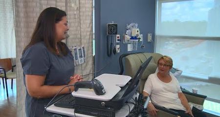
At the beginning of the surgery, the surgical team creates a “map” of the brain to preserve critical areas of functional anatomy. The patient is awake for this. Using an app called the NeuroMapper, the team tests a wide range of functions, and records patient responses. This app was developed by the Medical College of Wisconsin, in partnership with the University of Wisconsin Milwaukee. It gives surgeons a tool to monitor brain function during surgery.
Designed for operating room use on an iPad, the NeuroMapper app generates tests that the neuropsychologist uses to monitor the patient’s cognition during brain surgery to “map” the brain’s terrain. The goal of brain mapping during surgery is to locate important functional areas of the brain and avoid them so patients have less risk of developing cognitive changes from the surgery. To preserve important areas, surgeons wake patients during surgery and test their language and other functions. In the past, this involved bringing props such as photos into the operating room, which had some limitations. NeuroMapper allows the surgical team to test a wide range of functions and record patient responses. To determine if these responses predict outcomes, a research consortium of academic medical centers around the U.S. have put NeuroMapper to use during brain surgeries with the purpose of sharing collective data and yielding more systematic brain mapping protocols.
To start mapping, the surgeon first exposes the area of the brain where the tumor is located by making a small opening in the skull. Next, electrodes are placed directly on the brain to provide an accurate reading of brain waves. Different areas of the brain are then carefully stimulated with electrical current, which temporarily disrupts function.
The neuropsychologist asks the patient to do a variety of tasks, such as identifying pictures, clues and common words, and records the patient’s response. If the patient performed well during baseline testing but cannot accurately answer questions during surgery, the surgeon will adapt by taking a different path to remove tumor tissue. In this way, a neurosurgeon avoids areas that control important functions.
Most patients respond well to awake surgery because they can be part of their own treatment process, and the entire team works hard to keep them comfortable and relaxed. The neuropsychologist has guided imagery and other calming techniques at the ready to help avert anxiety if needed, and signals team members to address discomfort immediately.
Awake brain surgery requires a wide range of focused expertise, planning, coordination and cooperation on the patient’s behalf. Without this intricate multidisciplinary approach, awake surgery would not be possible.
-
Keyhole Surgery for Benign Brain Tumors
Keyhole surgery is a minimally invasive endoscopic technique used mainly for benign brain tumors. Using an endoscope, which is a slim instrument that has a tiny camera on the end, to guide the surgery, surgeons reach brain tumors through the nostrils and other access points on the skull including the corners of the eye, the creases of the eyebrow and behind the ear. When the tumor and its location demand creation of an opening, the surgeon performs a craniotomy, making an opening about the size of a quarter in the bone of the skull to accommodate the endoscope and surgical instruments.
The endoscope gives surgeons a precise, wide-angle view of the tumor and its margins, as well as the structures around it. Surgical instruments are also designed for long, narrow passageways. In addition, high-definition(HD) monitors in upgraded surgical suites aid the surgeon’s ability to view the tumor and avoid areas of the brain that control speech, memory and other vital cognitive functions. The surgeon can also use a dilator to access tumors deep inside the brain. After the tumor has been removed, if an opening in the skull was required, it is sealed with bone cement to prevent infection and brain fluid leaks.
Although tumor characteristics will dictate eligibility for keyhole surgery, people who may benefit from this surgery include those with meningiomas, schwannomas, pituitary tumors, pineal tumors, lesions that cause trigeminal neuralgia, and people who have metastases to the brain from cancer in other areas of the body.
MRI Surgical Suite and Neuro-Navigation System
A host of technologies and techniques are critical to successful brain tumor surgery. No matter the type of surgery, the common thread is image guidance. Magnetic resonance (MR) image guidance during surgery is the mainstay of operating on delicate structures like the brain that demand precise visualization throughout the entire surgery no matter the surgical approach.
At Froedtert Hospital, brain surgeries are performed in a special MR surgical suite that allows real-time access to MR guidance. With intraoperative MRI, complications are fewer, and second surgeries and tumor recurrence rates (due to not completely removing tumor tissue) are less likely. These are all critical factors in a patient’s recovery and outcome.
Neuro-navigational technology is another precision tool our neurosurgeons use to plan surgery and guide the surgical team during an operation performed in the MRI surgical suite. The team records the head’s position by translating MR images of strategically placed temporary markers on your scalp to a computer. A viewing device then projects this image to a monitor in the surgical suite.
By melding the image on the monitor with an MRI scan, the surgical team can see the head’s orientation at any time. They can also clearly view the tumor and its margins (edges that meet healthy tissue) so they can avoid healthy areas of the brain. Navigating the surgery with this level of accuracy enhances the neurosurgeon’s ability to remove the entire tumor through a smaller incision with the potential of a shorter surgery.
Personalized Brain Tumor Surgeries: No Two Patients Alike
Everyone has unique brain anatomy, and tumors present in various ways. Our multidisciplinary team of brain tumor experts carefully considers many factors before recommending the type of surgery that will be the most successful. Although you may have the same type of tumor as another person, it can be a different grade at diagnosis, inhabit a different location in the brain, and affect the ability to function to a greater or lesser degree.
Images from MR or CT scans, pathology results from biopsy and your overall health and medical condition are all part of the decision process. Regardless of the type of surgery, the team will access the tumor by performing a craniotomy — removing a small portion of bone from the skull to create an opening. The opening is typically kept as small as possible while allowing surgical instruments to pass through. A smaller opening aids in recovery from the surgery.
Virtual Visits Are Available
Safe and convenient virtual visits by video let you get the care you need via a mobile device, tablet or computer wherever you are. We’ll gather your medical records for you and get our experts’ input so we can offer treatment options without an in-person visit. To schedule a virtual visit, call 1-866-680-0505.
More to Explore


