 At the Eye Institute, physicians have access to a full range of the most sophisticated diagnostic technology available. All patient tests are conducted by trained ophthalmic technicians and test results are interpreted by specialty physicians, from which the results and recommendations are provided to referring physicians.
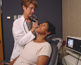
Ultrasound Images to Diagnose Eye Issues
At an eye exam, your physician can usually look through your pupil to see the back of the eye. However, changes in the eye sometimes can prevent a view into the back of the eye, or the physician may not be able to accurately measure the changes. When these situations occur, an ultrasound of the eyeball can be very helpful.
Ultrasound images and measurements help your physician diagnose changes in an eye and accurately measure eye structures for monitoring and treatment. An ultrasound of the eye involves numbing the eye and placing a small probe that admits sound waves on the numbed eye.
Patients feel a small vibration, though it is not painful. The reflection of these sound waves are then measured and processed to give an image of the structures in the back of the eye as well as accurate measurements of the changes. An eye ultrasound usually takes about one hour.
Ocular Photography for Glaucoma, Macular Degeneration and Other Eye Conditions
In treating any disease, it is important to keep exact records of changes in the condition over time. For eye diseases, often the best way to do this is with photographs of the inner structures of the eye. Ocular photography uses special equipment to capture images of the retina, the vitreous and the optic nerve. These photographs help physicians keep tabs on glaucoma, macular degeneration and other conditions.
First, the patient’s pupils are dilated with medicated eyedrops. Images of the eye are then captured using a special digital camera mounted on a microscope. The whole procedure takes about 10 minutes. Every procedure is performed by one of the Eye Institute’s ocular photographers.
Fluorescein Angiography to Diagnose Eye Disease
A key component to diagnosis and management of many eye diseases is to determine if there are abnormalities in blood vessels that line the back of the eye in the retina. Fluorescein Angiography (FA) allows for visualization of blood vessel and other abnormalities in the retina. This can be very helpful in the management of many ocular diseases such as diabetic retinopathy or age-related macular degeneration.
In this imaging procedure, a small amount of dye is injected into a vein in the patient’s arm. As the dye circulates through the blood vessels in the eye, pictures are taken so abnormal blood vessels and areas of blockage and leakage can be identified. These photos are very useful to physicians to help with diagnosis and guide treatment of many ocular diseases.
This imaging procedure is done through a dilated pupil. The fluorescein dye causes a brief harmless discoloration of the urine and skin for up to 24-48 hours. Adverse reactions are very rare but can include nausea, vomiting, and hives. Your eye doctor at the Eye Institute will be able to view your test within minutes of its completion.
Optical Coherence Tomography
Optical Coherence Tomography (OCT) is an imaging test that allows for detailed visualization of the different layers of the retina and the optic nerve. It helps physicians diagnose and monitor various diseases affecting the eye such as age-related macular degeneration and glaucoma.
The OCT image is captured by directing focused beams of light into the eye for a few seconds. These beams of light are reflected out of the eye and are processed to give very detailed cross-sectional images of the different structures of the eye. This test is non-invasive and takes about 15 minutes from start to finish. Eyes are usually dilated for this test.
Electrophysiology Tests
Occasionally a person experiences visual changes that are difficult to completely understand with only a clinical eye exam. These changes may be due to abnormalities in the function of some of the cells on the back of the eye in the retina, or there may be changes in the nerve connections between the eye and the brain.
Electrophysiological tests are types of tests that record responses from nerve cells in the back of the eye and can help your eye doctor better understand what is occurring in the eye.
For these types of tests, a contact lens with an electrode within it is placed on the eye. The patient then watches a screen with different lights to stimulate the retina. The electrical impulses from the retina are recorded. To get useful readings, the patient must first sit in the dark for a period of time to become completely dark-adapted. For this reason, this test usually takes about two hours.
