When it comes to uncovering the cause or extent of heart and vascular problems, all of the latest tools and techniques are available to our heart and vascular physicians.
As physicians examine each patient’s condition to pinpoint its cause and plan for treating the problem, they have access to many tests and procedures for diagnosing heart disease and vascular disease.
3-D echocardiogram — uses ultrasound waves to investigate the action of the heart, presented in a three-dimensional (3-D) image. We have the longest clinical experience in the state using this technique. Learn more about echocardiograms.
64-Slice LightSpeed Volume Computed Tomography (VCT) Scan — a powerful medical CT scanner that is unique in its ability to produce clear, three-dimensional images of blood vessels to detect coronary artery disease (CAD); VCT combines rapid X-ray scanning with multiple CT to produce highly detailed images of the heart and vessels. We were the first in the world to use the 64-Slice LightSpeed VCT Scan technology. Learn more about the 64-Slice LightSpeed VCT Scan.
Angiography — a procedure performed to view blood vessels after injecting them with a dye that outlines them on X-ray.
Ankle brachial index (ABI) — a simple, non-invasive, painless test that compares blood pressure readings in a person’s ankles with blood pressure readings in the arms. Learn more about ABI and other tests provided in our vascular laboratory.
Cardiac catheterization — the process of inserting a catheter into a vein or artery and guiding it through the heart chambers and surrounding vessels for purposes of examination or treatment. Learn more about cardiac catheterization.
Cardiac mapping, 3-D computerized electro-anatomical mapping — used to locate abnormal areas in the heart’s electrical system; electrode catheters are inserted into the heart to track the heart’s electrical signals. Our electrophysiologists use this sophisticated technique to visualize the heart and sources of irregular beats (arrhythmias). Learn more about our Arrhythmia Program for information about this test and other resources for patients with irregular heartbeats.
Cardiac MRI (cardiac magnetic resonance imaging) — the use of magnetic resonance imaging (MRI) to obtain images of the heart; MRI uses a magnetic field, radio waves and a computer to produce detailed pictures of internal body structures. Learn more about MRI.
Cardiac nuclear medicine imaging — uses radioactive materials (tracers) to obtain diagnostic images of specific parts of the heart and vessels. Two main types of these tests are multiple gated acquisition (MUGA) and nuclear medicine stress tests, also called myocardial perfusion imaging. Learn more about cardiac nuclear imaging.
Cardiac ultrasound — see echocardiogram
Carotid duplex scan — a non-invasive vascular ultrasound test to assess the blood flow of the arteries that supply blood from the heart through the neck to the brain. Learn more about duplex scanning and other tests provided in our vascular laboratory.
Cholesterol test, lipid panel — a commonly used blood test that measures levels of cholesterol, triglycerides, high-density lipoprotein (HDL) and low-density lipoprotein (LDL). Learn more about resources like cholesterol tests provided through our Preventive Cardiology Program.
Coronary artery calcium scan/scoring — a non-invasive test that uses CT imaging to measure the amount of calcium in the arteries. Also called a heart scan, it helps determine the risk of coronary artery disease (CAD) or heart attack.
CT angiography (CTA)/cardiac CTA — produces images of the blood vessels and tissues in a particular body structure by combining computerized tomography (CT) scanning with an injection of a special dye. A CT scan is a type of X-ray that uses a computer to make cross-sectional images of the body. Cardiac CTA produces images of the heart’s structure and function, including valves and chambers. We are leading the way in next-generation CT angiography with the 64-Slice LightSpeed Volume Computed Tomography (VCT) scanner, offering the most advanced, detailed images available. Learn more about CTA.
CT coronary angiography — a noninvasive alternative for cardiac catheterization that looks specifically at the blood vessels in the heart to diagnose coronary artery disease. Learn more about CT coronary angiograms.
Dobutamine stress echo test — an echocardiography test that involves taking a medication called dobutamine while being closely monitored; the medication stimulates the heart as if a person is exercising; the test is used to evaluate a person’s heart and valve function when he or she is unable to exercise on a treadmill or stationary cycle. Learn more about echocardiograms.
Drug stress test — a test that involves taking a medication while being closely monitored; the medication stimulates the heart as if a person is exercising; the test is used to evaluate a person’s heart and valve function when he or she is unable to exercise on a treadmill or stationary cycle
Duplex ultrasound — a procedure that combines Doppler flow and conventional imaging information to allow physicians to see the structure of blood vessels; duplex ultrasound shows how blood is flowing through the vessels and measures the speed of the flow of blood; it can also estimate the diameter of a blood vessel and the amount of obstruction (if any) in a blood vessel
Echocardiogram (“echo”) — a test that uses sound waves to create a moving picture of the heart; the picture is much more detailed than an X-ray image and involves no radiation exposure. Our state-of-the-art Echocardiography Laboratory performs thousands of echo procedures a year, including transthoracic echocardiogram (TTE), transesophageal echocardiogram (TEE), intracardiac echocardiogram (ICE), and cardiac resynchronization therapy (CRT) evaluation. Learn more about echocardiograms.
Electrocardiogram (EKG, ECG) — a non-invasive procedure for recording electrical changes in the heart. We perform about 30,000 electrocardiography procedures each year. Other types of EKGs include holter monitors, event recorders and loop recorders. Learn more about electrocardiograms.
Electrophysiology (EP) study — an invasive procedure that tests the heart's electrical system. EP studies may be performed to help evaluate the effectiveness of medications, to “map” or locate the point of origin of a dysrhythmia, or to gather other important details about the patient’s condition. Learn more about EP studies.
Exercise stress test — a screening tool to test the effect of exercise on the heart; also called heart stress test or treadmill stress test.
Extremity arterial mapping — uses non-invasive duplex ultrasound technology to examine blood flow in the arteries in the arms and legs. Learn more about extremity arterial mapping and other tests provided in our vascular laboratory.
Event recorder (or loop recorder) — a device that records irregular heart rhythm episodes over extended periods of time. Learn more about event recorders.
HeartFlow® Analysis — a noninvasive test to detect the presence of coronary artery disease. The first of its kind test provides a personalized, color-coded 3D model of the coronary arteries, allowing physicians to determine how narrowed arteries impact blood flow without an invasive procedure. The Froedtert & MCW health network is a national leader in utilizing HeartFlow Analysis, having performed more than 500 tests.
Heart stress test — a screening test used to diagnose the presence and extent of coronary artery disease (CAD) during exercise
Holter monitor — a device that records heart rhythms over 24-48 hours. Learn more about holter monitors.
Intracardiac echocardiography (ICE) — a type of echocardiography that provides ultrasound images from inside the heart muscle; a tiny ultrasound probe is mounted on the tip of a catheter so it can be moved through the blood vessels directly into the heart. Learn more about echocardiograms.
Intravascular ultrasound (IVUS) — a procedure in which a tiny ultrasound device is placed into the coronary artery to give a cross-sectional view from inside the artery; IVUS can aid in the selection and sizing of stents and balloons, and can show that a stent has been properly placed
Lipid panel — see cholesterol test
Multiple gated acquisition (MUGA) — a nuclear imaging test that measures how much blood the heart pumps or “ejects” with each contraction (called the ejection fraction) and how quickly that blood is ejected. Learn more about cardiac nuclear imaging.
Magnetic resonance imaging and magnetic resonance angiography (MRI/MRA) — cardiac MRI uses radio waves, a magnetic field and a computer to create highly advanced images of the heart. Magnetic resonance angiography is a type of MRI that looks specifically at the blood vessels. Learn more about MRI and MRA.
Myocardial perfusion imaging — a test that evaluates coronary arteries by determining changes in blood flow to the heart during exercise; also called nuclear medicine stress test. Learn more about cardiac nuclear imaging.
Nuclear medicine stress test — a test that evaluates coronary arteries by determining changes in blood flow to the heart during exercise; also called myocardial perfusion imaging. Learn more about cardiac nuclear imaging.
Stress echo/doppler exams — used specifically for evaluating valve disease, an echocardiogram is done before, during, and sometimes after the heart is stressed, usually through exercise. The Doppler exam, which creates images of the heart in motion, uses sounds waves to measure the direction, amount and speed of the heart’s blood flow. Learn more about echocardiograms.
Supine bicycle stress test — a stress test performed while a person, lying flat in bed, pedals a bike which is attached to the bed.
Tilt table test — a procedure to determine the cause of blood pressure drop and fainting; the patient is placed on a table which is tilted upward by degrees to a vertical position; blood pressure, pulse and symptoms are recorded with the patient in each position.
Transesophageal echocardiogram (TEE) — a test in which an ultrasound probe is placed close to the heart to provide clearer pictures. A tube with a small ultrasound probe on it is gently placed into a patient’s esophagus. TEE can help diagnose abnormalities of the heart, and show the size of the heart, how well it pumps and damage to heart tissue. Learn more about echocardiograms.
Transthoracic echocardiogram (TTE) — a test in which an ultrasound transducer, which emits sound waves, is placed on the chest in the area of the heart; a TTE may be done to detect a problem in a heart valve, determine the size and functioning of the ventricles, evaluate the heart after a stroke, check for fluid collection in and around the heart, and look for congenital defects of the heart. Learn more about echocardiograms.
Treadmill exercise test — a variety of heart and vascular tests can be conducted while a patient walks on a treadmill to assess the heart during exercise; also called heart stress test and exercise stress test.
Vascular and interventional radiology — the diagnosis and treatment of blocked blood vessels using minimally invasive image-guided procedures
Venous duplex ultrasound — combines Doppler (high-frequency sound waves) flow and conventional ultrasound imaging to assess the structure of blood vessels, how blood is flowing through the vessels and the speed at which blood flows. Learn more about venous duplex ultrasound and other tests provided in our vascular laboratory.
Our Cardiovascular Program continues to receive recognition as one of the top programs nationally. We are honored to provide high-quality, effective care for even the most high-risk patients.
-
Check Out Our Heart and Vascular Program Awards and Recognition
In its 2024 Specialty Excellence Awards, Healthgrades recognized Froedtert Hospital as one of America’s 50 Best Hospitals for Cardiac Surgery, one of America’s 100 Best Hospitals for Cardiac Care and one of America’s 100 Best Hospitals for Coronary Intervention, as well as other specialty achievements in various areas.

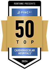
For the second year in a row, Froedtert Hospital was identified as one of the nation’s 50 Top Cardiovascular Hospitals™ according to an independent quality analysis based on a balanced scorecard provided by PINC AI™, and reported by Fortune. The hospitals recognized in the top 50 operated at lower cost and had better outcomes, recording significantly higher inpatient survival rates, fewer patients with complications, lower readmission rates and up to nearly $10,000 less in total costs per patient case. According to the study’s analysis, if all hospitals operated at the level of this year’s top performers, there could be 7,600 fewer deaths due to heart disease, 6,700 fewer bypass and angioplasty patients who suffer complications, and more than $1 billion in costs saved for the 2024 study year. Froedtert Hospital was ranked in the category of top teaching hospitals with a cardiovascular residency program. In this cohort of hospitals, Froedtert Hospital was ranked No. 4 in the country. No other hospital in Wisconsin was recognized with this national distinction.
The Society for Vascular Surgery's Vascular Quality Initiative (SVS VQI) has awarded Froedtert Hospital three out of three stars for its active participation in the Registry Participation Program. The mission of the SVS VQI is to improve patient safety and the quality of vascular care delivery by providing web-based collection, aggregation and analysis of clinical data submitted in registry format for all patients undergoing specific vascular treatments. The VQI operates 14 vascular registries.
The American Heart Association recognized Froedtert Hospital with its Get With the Guidelines® Heart Failure Gold Plus Award. In addition, the hospital was recognized on the AHA’s Target: Heart Failure(SM) Honor Roll and received the AHA’s Target: Type 2 Diabetes Honor Roll™ award.
The American Heart Association also recognized Froedtert Hospital with its Get With the Guidelines® — Coronary Artery Disease Mission: Lifeline STEMI Receiving Silver Plus and Mission: Lifeline NSTEMI Silver awards. These awards demonstrate our commitment to improving care by adhering to the latest treatment guidelines and streamlining processes to ensure timely and proper care for heart attacks.
The American Heart Association recognized Froedtert Hospital with its Get With the Guidelines® AFib Gold Award.
The Cardiovascular Intensive Care Unit (CVICU) and Neurosurgical Intensive Care Unit (NICU) at Froedtert Hospital have each received a silver-level Beacon Award for Excellence from the American Association of Critical-Care Nurses. This award recognizes unit caregivers who successfully improve patient outcomes and align practices with AACN’s six Healthy Work Environment Standards. Receiving this national three-year award with gold, silver and bronze designations, marks a significant milestone on the path to exceptional patient care and achieving a healthy work environment.


