Medical technology offers arrhythmia patients many options for implanting small devices that correct, monitor and restart the beating heart.
Pacemakers Help Maintain a Regular Heart Rhythm
A pacemaker is a small device that helps the heart beat in a regular rhythm. It uses batteries to send electrical impulses to the heart. There are different types of pacemakers for different needs.
In a minimally invasive surgical procedure (using local anesthesia), a small incision is made in the chest, where the pacemaker and leads are inserted. (A lead is a flexible wire that connects the pacemaker to the heart muscle.) The lead is attached to the heart and the other end is attached to a pulse generator placed in the chest (under the skin). After a pacemaker is implanted, it is programmed to the precise needs of the patient.
WATCHMAN FLX™ Device
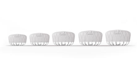
Patients with nonvalvular atrial fibrillation (AFib or AF) may be eligible for the WATCHMAN FLX™ device — an alternative treatment to long-term anticoagulation therapy with Coumadin® (warfarin) and other anticoagulants. The WATCHMAN FLX device, a left atrial appendage closure (LAAC) device, is available for implantation in eligible patients.
While anticoagulation therapy has proven an effective treatment for many patients with AFib, for some patients it is not well tolerated long term. The WATCHMAN FLX device can be an effective alternative treatment for these patients.
The WATCHMAN FLX device is the newest LAAC device, and is designed to advance procedural performance and safety while also expanding the population of patients that can be treated. Froedtert Hospital is one of only a select few in the region currently offering this new device to patients.


The WATCHMAN FLX LAAC device is inserted via a one-time minimally invasive procedure, and is designed to close the left atrial appendage (LAA) for many anatomies to prevent migration of blood clots, thus reducing the risk of stroke and systemic embolism.
The WATCHMAN device helped a local woman eliminate the need for warfarin. Read Linda's story.
Implantable Cardioverter Defibrillators (ICDs) Prevent Cardiac Arrest
An ICD continuously monitors the heart’s rhythm and recognizes rapid arrhythmias such as ventricular tachycardia (VT) and ventricular fibrillation (VF). The ICD is the most reliable way to protect against cardiac arrest and sudden death. The defibrillator corrects the rapid rhythm by delivering a high-voltage electrical shock, when needed, to restore a normal heartbeat. Less often, a person might have a heart rhythm that is too slow. In this case, a pacemaker (built into all ICDs) will send a low electrical pulse to the heart to “remind” it to beat.
An ICD is implanted during a minimally invasive surgical procedure.
Biventricular Devices for Cardiac Resynchronization Therapy (CRT)
Some patients will benefit from the implantation of a special pacemaker or defibrillator that stimulates the right and left ventricles to help them contract in sync and pump more effectively. These are called biventricular devices. Medical College of Wisconsin electrophysiologists are highly skilled in using biventricular devices in a technique called cardiac resynchronization therapy (CRT).
There are two types of bi-ventricular devices:
- Pacemakers (CRT-P)
- Defibrillators (CRT-D)
These are special variations of the basic devices that add a third heart lead, which goes through the veins to pace, or stimulate, the left side of the heart. In certain people with heart failure, pacing both sides of the heart at the same time can improve energy levels, decrease shortness of breath, and even prolong life.
The bi-ventricular device implant is performed similarly to the basic device.
Loop Recorders Help Diagnose Causes of Fainting
Implantable loop recorders are used to help pinpoint the cause of syncope (fainting), if the episodes are too far apart to be recorded by external monitors.
The small loop recorder device is inserted during a simple outpatient procedure under local anesthesia. It is placed just below the collar bone (usually on the patients left side). Similar to an electrocardiogram, the device continuously records heart activity for up to 2-3 years. If the patient experiences a fainting episode the device is activated to record the heart’s activity before, during, and after the episode. An electrophysiologist then evaluates the recordings to help determine the cause of fainting.
Virtual Visits Are Available
Safe and convenient virtual visits by video let you get the care you need via a mobile device, tablet or computer wherever you are. We'll assess your condition and develop a treatment plan right away. To schedule a virtual visit, call 414-777-7700.
More to Explore


