If the doctor detects an abnormality in your screening mammogram, you may be referred for a diagnostic mammogram or other specialized imaging. Radiologists at the Breast Care Center use several different imaging technologies to investigate breast abnormalities and gain additional knowledge about confirmed tumors:
Diagnostic Mammogram
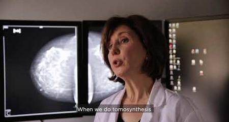
Mammogram technology can be used to investigate abnormalities detected through screening mammograms. A diagnostic mammogram captures images of the breast from additional views and angles.
Breast Ultrasound
Ultrasound is used to evaluate abnormalities detected through mammography or physical examination. For example, a mammogram might pick up a mass that appears to be a benign cyst. A breast ultrasound procedure can determine whether the mass is fluid-filled or solid, thereby confirming the benign diagnosis or indicating a need for further investigation.
Image-Guided Core Biopsy
Some breast abnormalities cannot be adequately evaluated through imaging studies alone. A sample of the abnormal tissue must be examined by a pathologist in a laboratory to make an accurate diagnosis. Image-guided core biopsy is a simple procedure in which a radiologist collects a small amount of breast tissue using a hollow biopsy needle. During the procedure, ultrasound or stereotactic (mammographic) guidance is used to position the needle at the site of the suspicious tissue.
Breast MRI
Many women who are newly diagnosed with breast cancer undergo a magnetic resonance imaging (MRI) study to determine the extent of their disease. MRI is very sensitive to differences in soft tissues, so it is able to detect some additional cancers that are missed by other imaging technologies. An MRI procedure can help detect smaller cancerous lesions that are adjacent to the diagnosed mass, elsewhere in the same breast or in the other breast.
Having an MRI is especially important for women with dense breast tissue. Breast MRI may also be used as a screening tool for women with a greater than 20 percent lifetime risk of developing breast cancer.
Image-Guided Localization
Women who will undergo surgery for a very small breast tumor (one that cannot be located easily by feel) first have a “localization” procedure. Using mammogram or ultrasound guidance, a radiologist places a small wire within the breast at the site of the cancerous lesion. During surgery, the wire allows surgeons to identify the tissues that need to be removed. For most patients, image-guided localization is performed immediately before surgery.
Mammograms and the COVID-19 Vaccine – What You Should Know
Swelling of the lymph nodes is a known side effect of the COVID-19 vaccine as well as other vaccines. Although it is temporary and not harmful, these enlarged lymph nodes may be seen on your mammogram. Because swollen lymph nodes can indicate breast cancer, we may call you back for additional evaluation and possible follow-up imaging.
Virtual Visits Are Available
Safe and convenient virtual visits by video let you get the care you need via a mobile device, tablet or computer wherever you are. We’ll gather your medical records for you and get our experts’ input so we can offer treatment options without an in-person visit. To schedule a virtual visit, call 1-866-680-0505.
More to Explore


