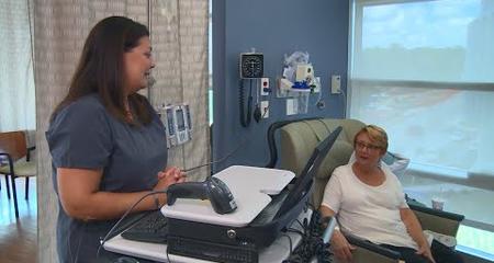
Optimal treatment results for patients with head and neck cancers happen when care is based on advanced diagnostic information. Physicians with Froedtert & the Medical College of Wisconsin have access to the full menu of tests to gather the diagnostic information they need to treat patients quickly and effectively.
An important first step after a thorough physical exam is to determine whether a tumor is cancerous or not. It is also vital to learn where it may have originated, as this knowledge impacts treatment recommendations.
State-of-the-Art Imaging (Radiology)
Radiology, or imaging, provides critical detail when diagnosing head, neck and skull base cancers. Imaging is used to help guide treatments, too. At Froedtert, our head and neck cancer specialists draw on a variety of imaging resources, including the following:
PET/CT
PET/CT combines the benefits of two powerful imaging approaches in one diagnostic scan. A positron emission tomography (PET) scan is a nuclear medicine study that detects the metabolism of a tumor, often revealing tumors that are hard to see with other types of imaging. A computed tomography (CT) scan uses high-definition X-rays to create thin, detailed cross-sectional images of the inside of the body. PET/CT quickly compiles the two scanning approaches to create a more comprehensive picture of the tumor and surrounding area. PET/CT scans also help physicians monitor how well a tumor responds to treatment.
64-slice Multidetector CT
The 64-slice multidetector CT, allows technicians to take very high quality cross-sectional images very quickly. Also known as LightSpeed VCT, or volume CT, this scan gives a finely detailed look at the anatomy of the head, neck and skull base. With the equipment’s 3-D computer capabilities, technicians produce images in many formats so that physicians can best plan surgical and radiation therapy treatments.
MRI
Magnetic resonance imaging (MRI) helps physicians see subtle differences in soft tissue, map a tumor and see if it is spreading along nerves. Froedtert Hospital has a 3 Tesla MRI scanner, the highest field strength scanner used in clinical practice.
Endoscopies and Biopsies
Biopsies involve removing a small tissue sample for examination under a microscope by a pathologist, a physician specially trained in identifying diseases. Tissue samples can be obtained in variety of ways. An endoscopy, when indicated, is often performed to explore internal areas such as the throat or voice box (laryngoscopy). An endoscopy uses a thin, flexible, tube-like instrument that has a light and lens for viewing and can be used for retrieving tissue samples, as well.
Sentinel Lymph Node Biopsy
In certain cases, we may perform a sentinel lymph node biopsy to determine if the cancer has spread to the lymph nodes. During a sentinel lymph node biopsy, a labeled dye is injected in and around the original cancer site. The dye is then transported by the body to the first lymph node (the “sentinel node”) to which cancer is likely to spread from that tumor. Your doctor will use a special imaging locating device to see the dye and identify the sentinel node or nodes (there can be more than one). The sentinel lymph node (or nodes) are then removed and tested to determine if the cancer has spread.
Virtual Visits Are Available
Safe and convenient virtual visits by video let you get the care you need via a mobile device, tablet or computer wherever you are. We’ll gather your medical records for you and get our experts’ input so we can offer treatment options without an in-person visit. To schedule a virtual visit, call 1-866-680-0505.
Recognized as High Performing by U.S. News & World Report
Froedtert Hospital is recognized by U.S. News & World Report as high performing in three adult specialties and 16 procedures and conditions, including cancer.


