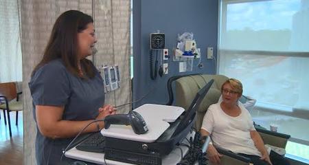
Regardless of the form it takes, any suspicious spot on the lung should be diagnosed quickly and accurately so an appropriate treatment plan can be developed. Early diagnosis usually translates into early treatment, which leads to better patient outcomes. There are a number of tests that can be used to diagnose a suspicious spot or mass on the lung, and each test may be performed by a different member of our team of specialists, including a thoracic surgeon, a radiologist or a pulmonologist. The tests conducted depend on each patient’s individual circumstances.
The diagnostic tool used depends on several factors, including the location and size of the suspicious spot or mass, the patient’s overall health and personal preferences, among other things. We tailor the test to the patient, as the first step in our comprehensive, multidisciplinary care. Which doctor performs the tests depends on a number of factors, and each specialist can perform more than one type of test. Our team works together closely, even during the diagnostic phase, to deliver the best possible care.
Tools to Diagnose Lung Cancer
Each diagnostic approach has its advantages and disadvantages, and all are based on evidence and research. We review each option thoroughly to help patients make the best choice for them. The tests and exams used for ruling out or diagnosing lung cancer include:
Physical Exam
A thorough physical exam and work up, including a complete medical history, is a key part of the diagnostic process.
Chest X-ray
A chest X-ray for another reason is often the reason a suspicious spot has been identified. X-ray is valuable in identifying masses that need further testing.
CT Scan
A CT scan can be used to identify abnormalities and to monitor changes or growth in a suspicious spot. We also use spiral CT, an advanced technology that uses a continuous spiraling motion to take detailed images quickly.
Bronchoscopy
This procedure uses a flexible bronchoscope, which is passed through the nose or the mouth, to see inside the lungs. It can be done under general anesthesia, or with sedation similar to that used during a colonoscopy. During a bronchoscopy, the doctor can remove tissue samples for biopsy.
Super Dimension® Bronchoscopy
 Also known as electromagnetic navigation bronchoscopy, this minimally invasive procedure allows the surgeon to biopsy lesions or masses from inside the lung rather than outside. It is especially effective in reaching lesions deep inside the lung and the lymph nodes in the area between the lungs, which may be out of reach of traditional bronchoscopes. Using a bronchoscope, computer-generated images and GPS-like technology, the physician can steer a catheter into the lesion to biopsy it.
This new technology is also useful for patients who might not be good candidates for a CT guided needle biopsy or a surgical biopsy. More and more patients can benefit from this procedure, which can have fewer complications than more invasive diagnostic procedures and is often more accurate, especially for lesions smaller than 2 cm.
CT Guided Needle Biopsy
This test uses CT imaging to guide a needle through the skin into the lung to remove cells or tissue from the suspected mass or lymph nodes for biopsy. It is most effective for tumors located very near the chest wall or the skin.
Video-Assisted Thoracoscopic Surgery (VATS)
This minimally invasive surgical approach uses a scope inserted between the ribs to examine the lung. Other small instruments can be inserted for the surgeon to perform any necessary diagnostic or therapeutic treatment procedures, including removing tissue for biopsy or removing the mass itself. VATS procedures typically result in less pain and faster recovery times than traditional open surgical approaches.
Endobronchial Ultrasound (EBUS)
EBUS uses an ultrasound probe at the end of a bronchoscope to help doctors see the mass they are biopsying to improve accuracy. It can be used to look at lymph nodes in the mediastinum (the area of the chest between the lungs) and a biopsy can also be obtained by passing a hollow needle through the bronchoscope.
Endoscopic Esophageal Ultrasound (EUS)
This technique uses an ultrasound probe at the end of an endoscope, which is passed down the throat into the esophagus. EUS allows doctors to see some deep lymph nodes and even the adrenal gland to determine if the cancer may have spread. A biopsy can also be obtained by passing a hollow needle through the endoscope.
Virtual Visits Are Available
Safe and convenient virtual visits by video let you get the care you need via a mobile device, tablet or computer wherever you are. We’ll gather your medical records for you and get our experts’ input so we can offer treatment options without an in-person visit. To schedule a virtual visit, call 1-866-680-0505.
More to Explore


